During the first year of life, the skull grows at a rate that has no parallel in later development. The brain approximately doubles in volume in the twelve months following birth, and the cranial vault expands in response. In a healthy infant, this growth proceeds along open sutures, producing a progressively rounder, more symmetric head shape. In an infant who has undergone craniosynostosis surgery, the same growth provides the biological force that post-operative helmet therapy is designed to direct — but the pace of change also means that any device designed to guide that growth has a limited window of relevance before the anatomy it was fitted to has moved on.
The infant had undergone craniosynostosis correction surgery. The procedure had released the fused suture; the work of guiding the skull toward improved symmetry now fell to post-operative cranial remoulding therapy. The treating neurosurgical team provided the clinical parameters — the target correction, the areas requiring preferential expansion, and the surfaces that should redirect rather than accommodate growth — and engaged Osteo3d to design and fabricate a custom helmet fitted to the infant's post-surgical head geometry.
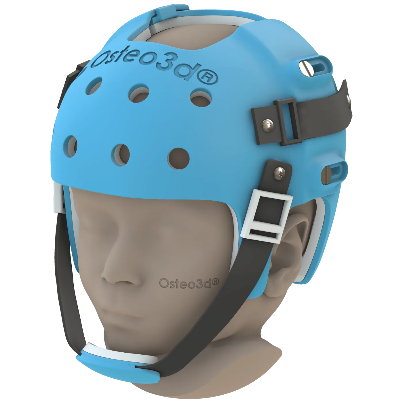
The first helmet was derived from the head geometry captured after surgery. The interior was designed with voids over the areas intended for preferential growth and contact surfaces elsewhere — the mechanical encoding of the correction target in a physical device the child would wear for the majority of each day. From the first fitting, the helmet directed growth rather than accommodating the residual asymmetry.
Rapid cranial expansion in early infancy means that a helmet fabricated at the start of therapy will not remain appropriately fitted throughout it. As the vault expands, the relationship between the helmet's interior and the skull surface shifts. At the point where that change was sufficient to reduce the effectiveness of the first device, a second helmet was fabricated — based on the updated head geometry and the correction still to be achieved. This sequential approach allowed the therapy to remain precisely targeted across the full six-month course rather than working from an increasingly imprecise fit.
Over the six months of therapy, the skull showed progressive improvement in contour and symmetry. The asymmetry that had remained after surgery was substantially reduced, and the cranial balance at the end of the helmet course was meaningfully better than at the start. The neurosurgical team was satisfied with the outcome of the post-operative phase.
The interaction between craniosynostosis surgery and post-operative helmet therapy is a clinical partnership: the surgeon creates the conditions for correction, and the helmet guides the correction that the biology provides. In rapidly growing infants, the precision of that partnership depends on devices that fit accurately at each stage of development. Sequential helmets, each designed from the actual head geometry at the point of transition, are a direct response to the pace at which early infant anatomy changes.
Osteo3d Team
Clinical Affairs