These papers are authored independently by the research teams named in them.
Springer2024
Mandibular Reconstruction Modalities Using Virtual Surgical Planning and 3D Printing Technology: A Tertiary Care Centre Experience
Clinical research team
Documents Osteo3d-assisted mandibular reconstruction across a tertiary care centre using virtual surgical planning and patient-specific 3D printed guides.
Digital Mapping of a Massive Skull-Base Ameloblastoma with Intracranial Extension, Resection, and Reconstruction Using 3D Templates and Molds: Descriptive Case Report and Review
Surgical and research team
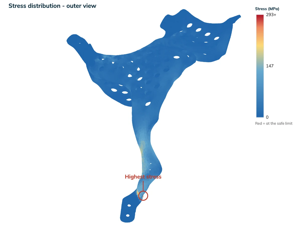
Osteo3d digitally mapped a massive skull-base ameloblastoma with intracranial extension, fabricated a patient-specific resection template encoding surgical margins, and produced custom 3D molds for PMMA bone cement reconstruction of the resulting craniofacial defect.
Customized and Cost-Effective 3D Printed Mold for Cranioplasty: India's First Single Center Experience
Neurosurgery team
Documents India's first single-centre experience using an indigenously developed 3D printed mold for cranioplasty, created using Osteo3d software and manufacturing.
Journal of Cleft Lip Palate and Craniofacial AnomaliesJul–Dec 2016
Case series of three-dimensional printing technology applied in complex craniofacial deformity surgery
Mendonca et al.
One of the earliest peer-reviewed publications featuring Osteo3d — discusses 3D-printed models used in complex craniofacial surgery at significantly reduced cost.
Used Osteo3d in published research and want the paper listed here? Get in touch.
Questions
Questions
Who is this for?
Clinical researchers, postgraduate candidates and academic departments who need imaging, 3D, simulation or measurement work they cannot do in house. It is for studies. A single patient goes through the clinical route at Solutions.
Do we need ethics committee approval before starting?
Approval, or a documented waiver, is needed before study data is accepted. The approval is held by you as the investigator. Protocol and imaging design can be discussed before approval is in place.
What is actually delivered?
A report, the figures your paper needs, and a private link to open and rotate the models in a browser. Physical models, where a study needs them, are printed by Osteo3d.
What data do you need, and in what form?
De-identified DICOM, one folder per subject, together with the question the study is answering. A data preparation checklist is available on the research brief page.
Who owns the results?
You own the study data, the measurements and the report, and you publish them as your own. Osteo3d retains its methods, its tooling, the models it builds, and any protocol researched during the work. This is set out in the engagement terms before anything begins.
Will Osteo3d be an author?
Only where the contribution meets the ICMJE authorship criteria, and only by agreement in writing beforehand. Otherwise an acknowledgement is supplied.
What does it cost?
Every engagement is quoted against its scope. Send a brief and a written scope with a price is returned.
What is not offered?
Manufacturing files of any kind, device design, manuscript writing, clinical interpretation of images, and software licences. Work on a single patient goes through the clinical route at Solutions rather than here. Reports produced here are for research use and are not a clinical decision aid.
Can Osteo3d supply data from its own archive?
No. Osteo3d holds clinical case data under the terms it was collected for, and it is not available for third-party research.
Send a research brief
One page describing the question, the imaging you hold and your target date. A written scope and a price come back.
We use analytics cookies to understand how visitors use this site. By clicking “Accept”, you consent to this under India's DPDP Act 2023. You may decline and the site will work without tracking.