The geometry of facial asymmetry presents a planning challenge that clinical examination and two-dimensional imaging cannot fully resolve. What is visible on the surface — a chin deviating from the midline, a lower jaw that sits off-centre, an occlusal plane that is canted — is the sum of skeletal contributions that cannot be separated without three-dimensional analysis. The mandible may deviate because of its own asymmetric growth, because the maxilla carries a canted occlusal plane, or because both have developed in compensation with each other. Which jaw to move, by how much, and in which direction cannot be reliably determined from a lateral cephalogram and dental models alone.
The patient presented with a clinically apparent skeletal asymmetry: deviation of the lower jaw from the facial midline, a displaced chin, and a malocclusion reflecting the underlying skeletal imbalance. The surgical team elected to address the deformity comprehensively — repositioning both jaws and correcting the chin — and requested virtual surgical planning to establish the precise movements required before committing to a surgical approach.
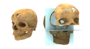
Using the patient's CT imaging and digital dental scan data, the bony anatomy was reconstructed in three dimensions and the asymmetry mapped quantitatively: the degree of mandibular deviation, the cant of the maxillary occlusal plane, and the chin displacement relative to the facial midline. The surgical movements were simulated digitally, with the target position confirmed by the surgical team before any fabrication began. As part of the workflow, 3D printed post-planning anatomical models were produced to represent the planned correction. These models served as a reference for the surgeons in the operating room, helping visualise the target alignment and symmetry to be achieved during surgery. From the finalised plan, surgical splints were fabricated to guide jaw positioning intraoperatively, and a patient-specific cutting guide was designed for the genioplasty — the chin repositioning component of the procedure.
In the operating room, the splints served their intended function: encoding the planned jaw position and transferring it to the surgical field. As is not uncommon with complex asymmetry cases, intraoperative splint positioning required some adjustment — the translation of a digital plan into the mechanical reality of bone, soft tissue, and field conditions rarely proceeds without adaptation. The surgical team managed these factors and completed the repositioning. The genioplasty cutting guide directed the chin osteotomy to the planned position.
The post-operative result showed clinically meaningful improvement in facial symmetry and occlusal relationship. The lower jaw and chin were repositioned toward the midline, the malocclusion was corrected to the planned target, and the patient's facial appearance — the primary concern driving the referral — improved substantially.
Facial asymmetry correction is technically among the more demanding applications of orthognathic surgery because the planning problem is genuinely three-dimensional in a way that simpler jaw advancement cases are not. Virtual surgical planning does not simplify the intraoperative work — surgical judgement and adaptation remain essential, as this case demonstrated. What it does is ensure that the target is clearly defined before the incision, that the team has agreed on what correction is achievable, and that the guides in the operating room represent the best pre-operative approximation of that target rather than an estimate made under time pressure.
Osteo3d Team
Clinical Affairs