An intraosseous tumour of the mandible presents a surgical problem that is, in one sense, two problems. The first is oncological: the affected bone must be removed with adequate margins. The second is reconstructive — the jaw that remains must function, and the geometry of the face must be restored. The difficulty of the second scales directly with the size of the first.
The patient presented with a large expansile lesion arising from within the mandible. Over time, the tumour had expanded to occupy a significant portion of the jaw, reaching approximately 7–8 cm in its greatest dimension. Lesions of this size distort the bony architecture substantially — the cortical walls thin, the inferior border remodels, and the relationship of the mandibular segments to one another shifts. By the time the patient reached surgical planning, the extent of involvement made straightforward resection and reconstruction a genuinely demanding undertaking.
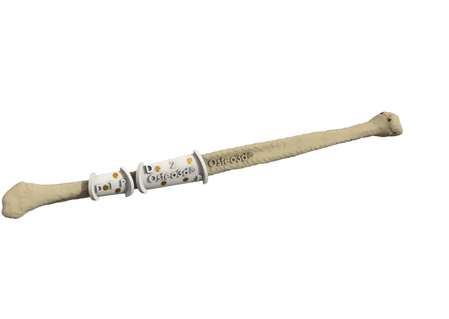
The surgical plan was a segmental mandibular resection followed by reconstruction with a fibula free flap. The fibula — taken from the lower leg along with its vascular pedicle — is the standard approach for mandibular reconstruction: it provides adequate bone length, tolerates multiple osteotomy cuts, and can be shaped to approximate the curved geometry of the jaw. But for a large resection involving a distorted mandible, shaping the fibula accurately demands pre-operative precision. The junction angles, the number of cuts, and the final positioning of the reconstructed segment must be planned — not estimated intraoperatively under time pressure.
Osteo3d supported the surgical team through virtual surgical planning using the patient's CT imaging. The mandibular anatomy, the extent of the resection, and the planned fibula configuration were worked through in the digital environment before the procedure. From that plan, patient-specific cutting guides were fabricated: one set to define the resection margins on the mandible, and a second to guide the osteotomies on the fibula. The guides encoded the geometry agreed upon in the plan — the angles, the segment lengths, the final contour — so that the reconstruction could be executed with precision rather than adjusted by eye in the operating room.
The procedure was carried out with the cutting guides in use. The tumour-bearing segment was removed, the fibula was shaped to the planned configuration, and the reconstruction was completed with the flap surviving. Given the extent of the resection, the recovery period was longer than a routine reconstruction. With time and rehabilitation, the patient regained functional jaw movement and the ability to eat and speak normally. The surgical team subsequently began planning for dental rehabilitation — the next phase of restoring full oral function.
Cases of this scale illustrate the point at which pre-operative planning shifts from useful to essential. A fibula free flap can be contoured freehand for a small defect with acceptable results. For a large reconstruction involving a distorted mandible — where the surgeon must recreate several centimetres of complex three-dimensional geometry while managing ischaemia time — the planning done before the incision determines what is achievable inside the operating room.
Osteo3d Team
Clinical Affairs